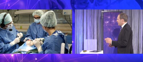
The Live Surgery Day, staged by the EFP and the Osteology Foundation on June 17, was hailed as a “very successful experience” by Mariano Sanz, one of the event’s moderators.
He praised the structure of the online event, which was attended by more than 1,000 people from more than 80 countries. “The format of a background keynote lecture, a live surgery, and a panel discussion with experts turned out to be very entertaining, dynamic, and inspirational,” said Prof Sanz.
“I am sure that the high quality of the lectures and the superb performance of the surgeons helped the participants not only to learn how to indicate, organise, and execute the surgical interventions but also provided them with hints and tricks that are fully applicable to everyday clinical practice.”
The Live Surgery Day was introduced by the presidents of the two organisations, William Giannobile for the Osteology Foundation and Lior Shapira for the EFP.
The first session, moderated by Mariano Sanz, covered recession in the mandible and featured a presentation by Martina Stefanini on the possibilities and limitations of this kind of surgery, which is often complex because of the unfavourable anatomical position.
She discussed the role of factors such as tooth position, vestibulum depth, and tissue phenotype and the various techniques that can be deployed – both classic techniques (free gingival graft, the modified two-step approach, and the laterally and coronally moved flap) and the newer procedures of the vertically coronally advanced flap (VCAT) and the laterally closed tunnel (LCT).
Former EFP president Anton Sculean then performed surgery on a patient with an RT2 (Miller class III) recession in the lower right central incisor 41 and with a thin phenotype. The objective of the surgery was to improve oral hygiene, alleviate pain, and improve aesthetics and the technique used was the LCT, which offers advantages in cases of a thin phenotype with little or no attached gingiva.
The second session, moderated by Givoanni Salvi, featured a presentation by Frank Schwarz on surgical techniques for peri-implantitis surgery in the anterior zone in which he explained how the selection of the approach depends on the type of defect. He recommended a non-reconstructive approach for implants with a machined surface and a reconstructive approach in cases of class 1 defects where four walls are present. For more challenging cases – in fact, those most often encountered by clinicians – he proposed a combined approach.
EFP president-elect Andreas Stavropoulos then performed live surgery on a 34-year-old female patient with peri-implantitis around one of two implants and with a mesial defect with an infrabony component visible in the radiograph and with the presence of buccal bone dehiscence. The surgery performed involved surface cleaning with an air-polishing device, implantoplasty on the buccal side of the implant, the harvesting of autologous bone chips to fill the defect and covering it with a collagen membrane.
At the end of each session there were panel discussions with many interesting questions raised by participants. Topics covered included how verticality is restored with both the VCAP and LCT techniques, wound stability, whether implants should be placed in high-risk patients with a history of periodontitis, the influence of the implant surface on peri-implantitis progression, and which factors can help minimise the risk of peri-implantitis.


